Recently we did our first practical, which involved walking around the large lab, having a look at some dead (and live) specimens, and having a go at some biological drawing. Biological drawing is, in a way, easier than normal, artistic drawing, because it only requires features that help in the process of identifying the organism. The rules around how you draw the organism, however, are much more strict. For example, the brief guide we were given says that artistic shading is NEVER allowed.
I looked a bit weird dunking my camera into the anemone tank, but I managed to get some quite good photos in my opinion. I've also posted one of my biological drawings. There are a lot of photos, but I'll do my best to explain them!
All these blue photos were taken from outside the tropical coral tank in the reception area, and it's a small exhibition of coral fluorescence under UV (which is why the tank looks blue)
It took me quite a while to get a close up of the clownfish, as they moved quite quickly, and this is perhaps the best one. They look quite grumpy, which makes me a little more fond of them.
By far my favourite photograph of all of the ones I took that day is this one. This is a young starfish, and it is only about 2-3mm across, which just shows you how good the macro setting can be now!
I got some strange looks taking these pictures, and someone even said "I do hope that's waterproof". Nevertheless, dunking an expensive camera into water is still unnerving.
This is a close up of a sponge. A lot of people know that bath sponges used to be made of actual sponges, but not many people know that to get them into a state so that you can use them, the fishermen had to beat the sponges on rocks to break up and wash out the spicules. More on spicules later.
This belongs to the phylum sipuncula. I Did a biological drawing of this, which I shall post here later
These are the spicules taken from a sponge. This photo was taken through a microscope eyepiece, which required patience and a steady hand! Spicules basically make up the skeleton of the sponge, though are not actually bones (which is why it's not an invertebrate). You don't want to use a sponge as a sponge if it still has its spicules, they're like shards of glass. Speaking of glass, spicules are usually made out calcium compounds, but in the rare deep sea sponges, they are silicates because calcium is dissolved much easier below 1500m. So deep sea sponges grow glass shards. How terrifying.
This is the "skeleton" of a sponge, with all its spicules in position. Below, you can see a close-up of this, but you still can't properly see the individual spicules. I love the pattern they make.
More spicules. As you can see, their shape varies quite a bit.
A close-up of coral. I do think their structure is really quite beautiful.
A "forest" of coral. This skeleton is what is left in bleached coral- when the surrounding water gets too hot for the coral, which then expels the beneficial algae (which gives coral their spectacular colours). The coral cannot live without the algae, so it dies, and its white skeleton is left.
This is an interesting story. This used to be feared by sailors because it resembled the bloated hands and feet of dead children (nice image). It's actually a sponge that grows on the shells of hermit crabs, and gradually dissolves their shell until it becomes their shell. This actual specimen had a large hermit crab in it once.
And here's the photo of said hermit crab, when it was alive!
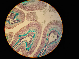
This is a slice of Anemonia viridis, which is obviously an anemone. Below I have posted the sort of thing we were asked to do.
I quite like this one because of the blue bits, which to my understanding are part of the anemone's collar, retracted into bundles so the tentacles are out.
This is a close-up of the anemone's mouth. I'd never seen its mouth before, so this was particularly interesting. It didn't really do much aside wiggling the tentacle you see near its mouth.
I have yet to find out what this actually is. It's one of the strangest things I've seen in the reception tank, as each blobby thing looks like a lobster eye. The colours were amazing, too.
That's it for today, I hope you enjoyed the pictures. I have been told I have an invertebrates practical next week, too, so I'll bring my camera along to that too. The workload is slowly increasing, but I think I'm managing to keep on top of it. I enjoy the information in some lectures so much sometimes that we have discussions on the bus home while we wait for the freight train to cross.
I love it here, I feel like I am in my element! (But I am looking forward to coming home for Christmas. I miss my double bed). Anyway that's really it. I'll post sometime next week, as I've worked out the posting weekly works best for me. So long!


Next update please.
ReplyDelete